Cells, Organelles: Basic and Acid Stains
The first step in preparing a tissue or organ for microscopic examination is fixation, or preservation, of the specimen. Formalin is a commonly used fixative. Many other fixatives are available and are used in the study of specific structures.
Note: There is a more complete description of methods for preparation of histological samples at the end of this laboratory manual (p. 92) under the heading "Histological Techniques".
The specimen on the microscope slide is a thin section (usually 5 micrometers) of the fixed tissue or organ. The section is stained by one or more dyes. Without staining the section would be nearly invisible with the microscope. Components of the specimen generally stain selectively and, on this basis, various regions of the specimen may be differentiated from each other.
Most dyes are neutral salts. In some stains, the dye moiety is a cation and such dyes are called cationic or basic dyes. These form salts with tissue anions, especially the phosphate groups of the nucleic acids and the sulfate groups of the glycosaminoglycans. When the dye moiety is an anion, the dye is called anionic or acid dye and salt formation occurs with tissue cations including the lysine and arginine groups of tissue proteins. Tissue components that recognize basic dyes are "basophilic" and those that recognize acid dyes are "acidophilic".
A common combination of stains is hematoxylin and eosin (H&E), which are commonly referred to as basic and acid dyes, respectively.
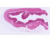
#39 Colon. H&E
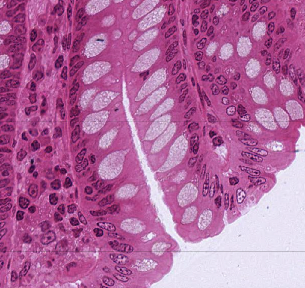
 This slide illustrates different kinds of cells; do not be concerned at this time with the structure of the colon. Large numbers of cells are seen. Nuclei are basophilic and are stained blue. At lower magnifications they appear as blue dots and at higher magnifications chromatin and nucleoli may be identified within the nucleus. Surrounding the nucleus is the acidophilic cytoplasm stained pink (due to the positive charges on arginine and lysine). The luminal surface (center of the slide) is smooth and consists of pale cells (called Goblet cells), absorptive cells, and enteroendocrine cells that make up the mucosa. These cells are forming columnar structures called intestinal glands. The free surface of the cell, facing a lumen, is referred to as the cell apex and the opposite surface is the cell base. The lateral borders should be seen and contain structures that connect the cells together. Note that (luminal) mucosa is densely packed with cells arranged in rows.
