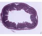
Simple Cuboidal Epithelia and Simple Columnar Epithelia
**Simple cuboidal epithelia
**
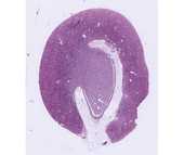
h3. #50 Kidney, H&E
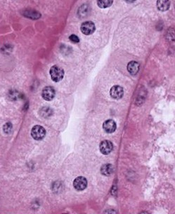 Examine the cuboidal epithelium that makes up the kidney tubules. Find the basement membrane and lumen of the tubules to help you determine the basal and apical membranes, respectively. Note that in some cases the lateral borders of cells are distinct while in many they are not. This is because they are highly interdigitated, a configuration that increases the surface area for transport across the cell membranes. This can be seen in electron micrographs of kidney tubules. Remember that each nucleus corresponds to one cell.
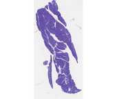
#107 Pancreas, Acid fuchsin and toluidine blue
Examine the epithelial cells. Note the basophilic structures that at the base of the cells are rough endoplasmic reticulum. At the apex of the cell, secretory granules appear as acidophilic structures. The contents of these granules are proteins, which are the precursors of digestive enzymes. Review the subcellular structures involved in protein synthesis.
Simple columnar epithelia
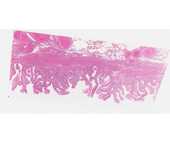
#115 Gall bladder. H&E
Examine the lining of the gall bladder as an example of simple columnar epithelium. Be sure you locate regions where the epithelium is cut longitudinally to observe the simple columnar epithelium. Note the microvillous border that you identified in the previous lab. In tangential sections portions of cells in various planes of section may give the impression that the epithelium is stratified. Why do the nuclei appear at different levels in tangential sections?
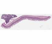
#101 Small Intestine (PAS and hematoxylin)
Locate the epithelium with its microvillous border and PAS positive glycocalyx. The basal lamina is also PAS positive, but is not intensely stained. What is the basis for PAS stain?
#102 Small Intestine (Bodian/silver)
 This slide illustrates modifications found in the apical region of the columnar epithelium: the striated (microvillous) border and the junctional complex. In longitudinally sectioned cells, the junctional complex is seen as a dark dot of silver deposit at the apical lateral borders of the cells. In regions where the epithelium has been cut in cross or oblique section, the junctional complex has a belt-like appearance and can be seen to encircle the cells (hexagonal shape). What types of intercellular junctions are commonly found in epithelia? Review the appearance of junctional complexes at the EM level in the electron micrograph in the lab.
In addition the Bodian silver stains secretory granules within enteroendocrine cells in the epithelium and the basal lamina.
