Adipose Connective Tissue
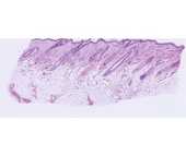
#46 Skin, scalp, H&E
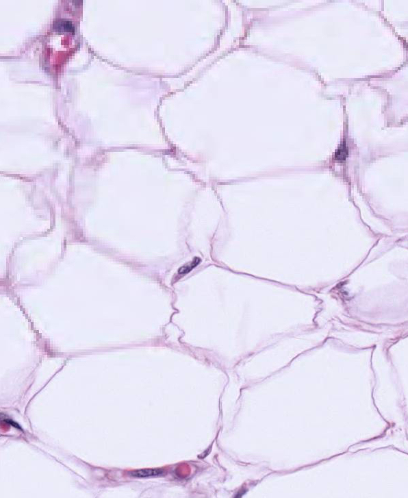
 Lying deep to the dermis is the loose subcutaneous connective tissue layer (superficial fascia). The subcutaneous connective tissue may be composed largely of adipose tissue. The epithelium (epidermis) and dense, irregularly arranged connective tissue appear deeply stained. The adipose connective is the palely stained region. At higher magnification observe that the intracytoplasmic lipid has been extracted from the fat cells during the histological preparation of the tissue. The thin peripheral ring of cytoplasm and the flattened peripheral nucleus, coupled with the large central vacuole results in the "signet ring" appearance of fat cells. In white fat each cell contains a single fat droplet (unilocular).
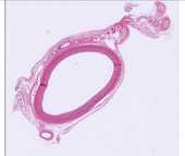
#16 Aorta, Cross Section
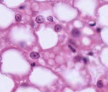
 In the connective tissue surrounding the aorta, note the presence of both white and brown adipose cells. At higher magnification observe the white fat in which each cell contains a single fat droplet (unilocular). In brown fat cells the lipid is accumulated in droplets, giving the cells a multilocular appearance. Where is the majority of brown fat found in humans?

