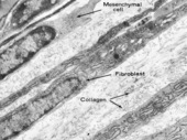
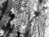
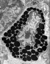
Electron Micrographs

Examine the electron Micrographs so that you understand the ultrastructural equivalents of the structures you have seen under the microscope.

Fibroblasts in Developing Connective Tissue

Active fibroblasts forming collagen have abundant RER and distended cisternae. The relatively undifferentiated cell (mesenchymal cell) at the upper left has a different chromatin pattern and little differentiation of its cytoplasm. The newly formed collagen fibrils are slender and more or less randomly oriented.
Bloom, W and Fawcett, DW, A Textbook of Histology 10th ed., WB Saunders, Philadelphia, 1975, p. 173.

Connective Tissue Matrix

Ground substance is fine granular material that fills the spaces between the collagen © (cut in cross and longitudinal section) and elastic (E) fibers (irregular electron dense objects) and surrounds fibroblast cells and processes (F). The granularity of the ground substance is an artifact of glutaraldehyde fixation.
Junqueira, LC and Carneiro, J, Basic Histology 11th ed., McGraw-Hill, New York, 2005. p. 112.

Mast Cell

This is a fully differentiated mast cell with a fairly heterochromatic nucleus. Numerous granules (Gr) containing histamine and heparin nearly fill the cytoplasm in which there are also a few mitochondria (M), free ribosomes ® and profiles of endoplasmic reticulum. There is no basal lamina around this wandering cell. At the left is a lymphocyte (Lc). (submucosa of rat gut)
Porter, KR and Bonneville, MA, Fine Structure of Cells and Tissues, 4th ed., Lea & Febiger, Philadelphia, 1973, p. 134.