Bone
Bone is a calcified connective tissue, and like other connective tissues, it consists of cells, fibers, and ground substance. The deposition of inorganic calcium phosphate salts as hydroxyapatite crystals within its matrix is a distinguishing characteristic of bone. This renders it structurally rigid. In addition, bone functions as a homeostatic reservoir of calcium and phosphate ions, and it encloses the hematopoietic elements of the bone marrow.
There are two types of mature bone - compact (lamellar) and spongy (trabecular or cancellous). Compact bone is characterized by the regularity of its collagen fibers. Spongy bone consists of a lattice of branching bony spicules, known as trabeculae, which are surrounded by bone marrow in some regions. When the trabeculae are sufficiently thick, they may contain osteons (see description below).
Immature (woven) bone: (see in "bone development") is the first bone laid down in prenatal life or in the repair of bone fractures. In this type of bone, the matrix immediately surrounding the osteoblast is called osteoid and is not mineralized. Immature bone is characterized by irregularly arranged, interwoven collagenous fibers within a matrix containing proteoglycans.
Because of its calcified matrix, bone presents difficulties in its preparation for microscopic study. There are two basic techniques for studying bone with the light microscope, and both of these types of preparations must be studied to appreciate the organic and inorganic components of bone. (1) Bone may be decalcified by acid solutions prior to embedding and sectioning. This permits study of the cells and organic matrix of the bone. (2) To study the lamellar and canalicular pattern of the calcified matrix, it is necessary to grind down dried bone that has not been decalcified to a thickness that permits the microscope light to be transmitted ("ground bone").
Ground Bone
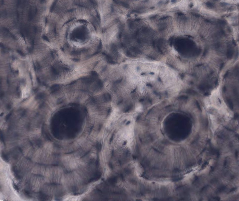
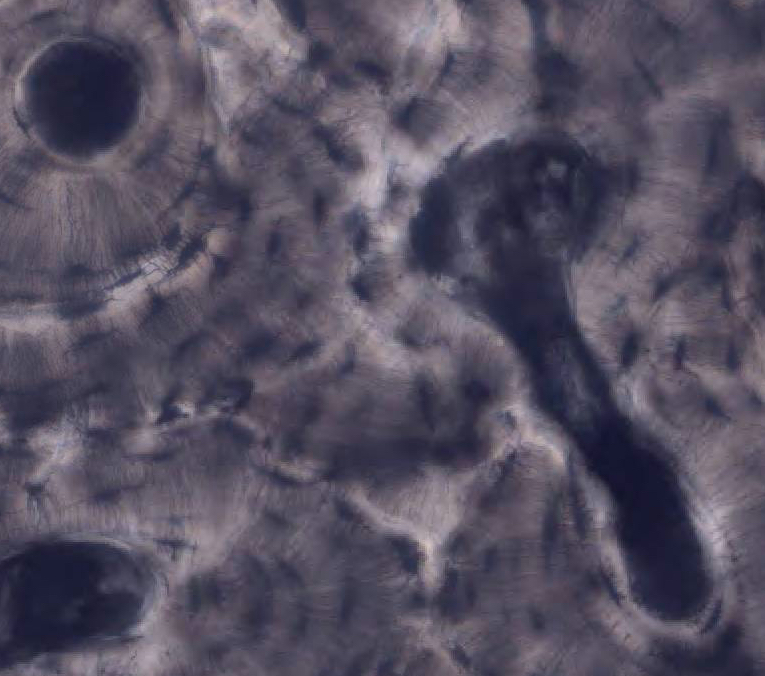
#9 Dried Bone, Shaft of Tibia
Cross and longitudinal sections (unstained). Use the illustrations in your textbook as a guide and identify the following structures.
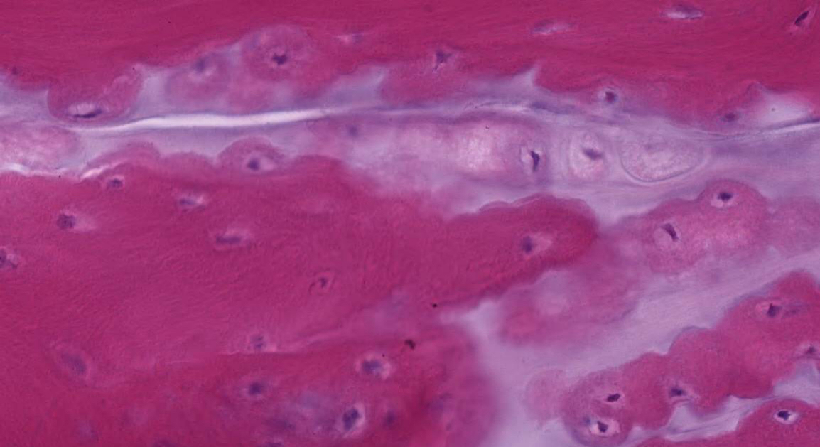
Haversian Systems (osteons) are distinctive structural units of compact bone that reflect the developmental and nutritive pattern of its lamellar configuration. Haversian systems consist of Haversian canals containing blood vessels and nerves surrounded by concentric lamellae of bone. Lacunae lie between or within the lamellae. In life these lacunae are occupied by osteocytes. Lacunae are connected with each other, and ultimately with the perivascular spaces of the Haversian canal, by canaliculi. This communicating system of canaliculi is essential for exchange of gases and metabolites between the osteocytes and the perivascular spaces of the Haversian canal. Volkmann's canals, which also contain vessel and nerves, are larger in diameter than Haversian canals and run perpendicularly to them.
Interstitial lamellae lie between the more distinct Haversian systems; these are the remnants of earlier Haversian systems that have been partially resorbed during bone remodeling.
Decalcified Bone
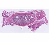
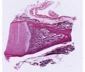
#11 Rib, Cross Section, (H&E).
 Surrounding the sectioned rib are bundles of skeletal muscle, tendon, nerves and blood vessels. Note the gradation of the surrounding connective tissue with the periosteum and the increased cellularity of the periosteum. The reversal lines (also known as cementing lines) that delimit the Haversian systems may appear refractile or slightly basophilic. What accounts for this basophilia? Trabeculae of bone extend into and partially subdivide the marrow cavity, which contains hematopoietic bone marrow. Bone marrow will be studied in more detail in a later lab.
#8 Rib and Cartilage, (H&E)
This slide demonstrates periosteum, which has dense cortical bone on the surface (better illustrated in the preceding slide) and spongy bone centrally. Osteoblasts are prominent on the surface of the bony trabeculae. Osteoclasts (multinucleated giant cells with acidophilic cytoplasm, related to the process of bone resorption) may also be seen near the osteochondral junction. Calcifying cartilage and rows of hyaline cartilage cells are present and extend into the cartilage of the proximal end of rib. Around the rib section, skeletal muscle and tendon are present.
Questions
- What structures are found within Haversian canals?
- Is the osseous lamella adjacent to the Haversian canal the youngest or the oldest lamella of a particular osteon?
Be sure you know how cartilage and bone differ morphologically, functionally, and with respect to blood supply.