Endochondral Ossification

Events in development of long bone:
- A hollow cylinder called the periosteal collar forms through intramembranous ossification around the middle of the cartilage model. The periosteal collar causes the underlying cartilage cells to begin to degenerate and die.
- The primary center of ossification begins by calcification of matrix of the diaphysis and eroding by blood vessels. These blood vessels bring osteoprogenitor cells with them when they penetrate the bone collar.
- The osteoprogentior cells differentiate into osteoblasts and begin depositing matrix, forming spicules.
- Secondary centers of ossification begin in the epiphysis at each end with invasion by blood vessels.
Growth at the epiphyseal plate:
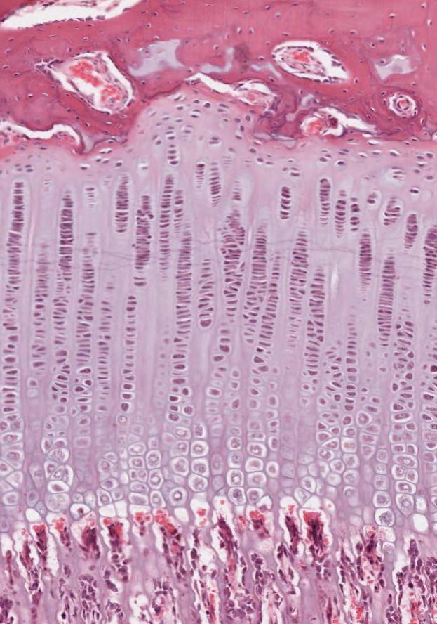
Passing from the articular end of the cartilage toward the ossification center of the diaphysis, the following zones are encountered in succession in the growth plate:
 # zone of reserve cells: A thin layer (3-6 cells wide) of small, randomly oriented chondrocytes adjacent to the bony trabeculae on the articular side of the growth plate.
# zone of proliferation: Chrondrocytes are stacked in prominent rows and the cartilage matrix becomes more basophilic in this zone. Mitotic figures are present and the axis of the mitotic figure usually is perpendicular to that of the row of chondrocytes.
# zone of hypertrophy: Chrondrocytes and their lacunae increase in size.
# zone of calcification: Deposition of minerals in the matrix surrounding the enlarged lacunae causing cell death.
# zone of ossification: Osteoblasts deposit bone matrix on the exposed plates of calcified cartilage.
# zone of resorption: Osteoclasts absorb the oldest ends of the bone spicules.
Note the high vascular density in this area: one capillary loop for each chondrocytic column. Narrow partitions of calcified cartilage are left behind as the bone grows in length.
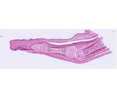
#97 Finger, human, 2 mos. H&E
This is a longitudinal section cut through an interphalangeal joint. Locate the ends of two long bones participating in the joint and identify the articular cartilage. Identify the epiphyseal disk, the metaphysis, the marrow cavity, and the diaphyseal bone. In the epiphyses where growth in length is occurring, note the zones of reserve cells, proliferation, maturation, hypertrophy, calcification, ossification and resorption. What structure in mature bone is created by the zone of resportion?
Each of these bones has a primary center of ossification. The zone of endochondral ossification spreads from the primary ossification center toward the ends of the cartilage. These slides do not show secondary ossification centers.
Note the bone of the diaphysis. Recall that this bone is growing in width by apposition and remodeling along the periosteum and the endosteum. In the marrow cavity note the bony spicules with calcified cartilage cores.
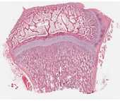
#96 Epiphyseal growth plate, H&E
As the primary ossification center of the diaphysis advances toward the epiphyses, each epiphyseal cartilage continues to grow and the whole cartilage model increases in length. This increase in length and extension of the primary ossification center results in a sequence of changes in the chondrocytes of the epiphyses, which is similar to that described for the establishment of the primary center.
In the epiphyseal growth plate, observe the zones of reserve cells, proliferation, maturation, hypertrophy, calcification, ossification and resorption.
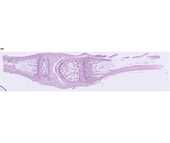
#95 Finger, monkey, 4 mos. H&E
Secondary ossification centers have developed in the epiphyses. Enlargement of the epiphysis occurs by growth of the articular cartilage. When growth ceases, the epiphyseal disk is entirely replaced by spongy bone and marrow ("closure of the epiphyses"), resulting in a visible epiphyseal line.
This slide includes a diarthrodial joint. In synovial or diarthrodial joints, articular cartilage caps the ends of the bones, which are kept apart by a synovial cavity filled with synovial fluid. The articulation is enclosed by a dense fibrous capsule, which is continuous with the periosteum over the bones. Internal to this is the synovium, a secretory membrane formed by a layer of collagenous fibers interspersed with flattened fibroblasts (synovial cells). This membrane is commonly thrown into folds (synovial villi) that project into the synovial cavity.


