Skeletal Muscle
Skeletal muscle is composed of large, cylindrical, multinucleated cells. The most striking feature of skeletal muscle fibers is the presence of striations, which are visible in longitudinally sectioned fibers. The striations are due to the presence of myofibrils, which are cylindrical bundles of thick and thin myofilaments, organized into units of contraction called sarcomeres. The orderly arrangement of these repeating units within the myofibrils gives rise to the characteristic pattern of transverse banding.
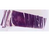
#15 Skeletal muscle (Phosphotungstic Acid - Hematoxylin, PTAH)
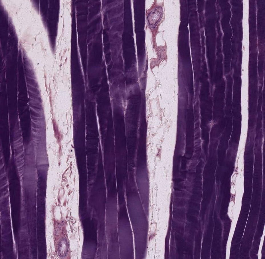 Examine this PTAH-stained preparation at low power and note that the muscle fibers are grouped into bundles (or fascicles). The spaces between the bundles contain the perimysium, in which connective tissue fibers, adipose cells, blood vessels and nerves can be identified under higher magnification. Next, under moderately high magnification, examine the individual skeletal muscle fibers, which have been cut lengthwise. Each fiber is separated by a delicate loose connective tissue (endomysium). Under higher magnification note myofibrillar cross-banding (alternating dark or A bands and light or I bands). Lastly, under highest power, look for additional markings, such as a thin Z band bisecting each I band and, in occasional fibers, a light H band within each A band. These distinctions may not be evident with light microscopy, but can easily be seen in TEMs (transmission electron micrographs). Note the shape and position of the pale-staining muscle nuclei, which should not be confused with the flattened, more elongate fibrocyte nuclei within the neighboring endo- or perimysium.
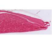
#3 Muscle and tendon junction, H&E
At low power examine bundles of skeletal muscle fibers and sheets of tendon (dense regular connective tissue). Under higher magnification, skeletal muscle fibers may be recognized by their cross-striations. In this preparation most tendon has a homogenous, almost glassy, appearance (this is a diagnostic feature). The cells of the tendon (fibroblasts or fibrocytes) occur in rows, squeezed between the thick collagenous fibers; only their flattened, rod-like basophilic nuclei show well. The zone of insertion of the skeletal muscle into the tendon is obvious, but higher resolution with the electron microscope is necessary to see the detailed structure of the junction.
Define a sarcomere. Be sure you know what the electron microscope has revealed about its fine structure. Know the structural changes that occur in a sarcomere during contraction and the theory that has evolved from electron microscopic studies to explain muscle contraction.
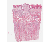
#116 Tongue, H&E
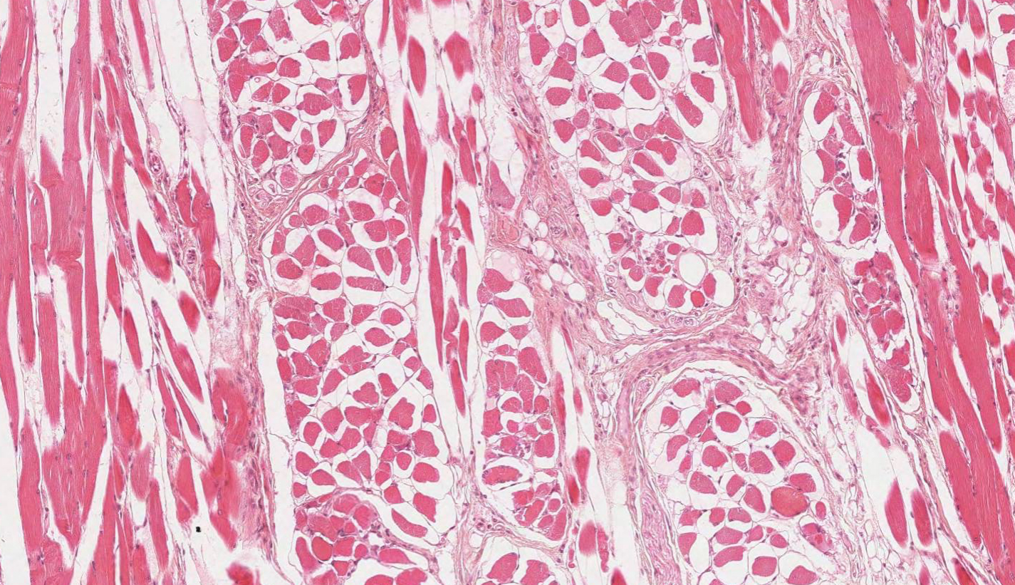 Observe numerous bundles (fascicles) of skeletal muscle fibers cut in different planes (this is a unique feature of the tongue). Identify the fatty connective tissue (perimysium), which surrounds each muscle bundle. From the perimysium partitions of loose connective tissue (endomysium) can be seen to penetrate into the bundle separating the individual muscle fibers. Under higher magnification, examine some skeletal muscle fibers in longitudinal section; striations may show poorly (if at all). Nuclei are clearly visible at the periphery of muscle fibers cut in cross-section; in fibers sectioned lengthwise, they may appear to occupy any position with respect to fiber breadth.
