Smooth Muscle
Smooth muscle is widely distributed in the body. It is found in the walls of ducts and blood and lymphatic vessels, as well as in the walls of the digestive, respiratory and urogenital tracts. It also occurs in many other sites including the eye (iris and ciliary body), skin (arrector pili muscles of hairs), endocardium, scrotum, penis, perineum, and nipple. Always correlate the function of smooth muscle with the different organs and regions in which it is found.
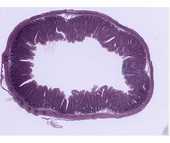
#39 Colon, cross section, H&E
 At low power one can easily distinguish the regions of the epithelium and connective tissue from that of the smooth muscle. Examine the outermost part of the pink acidophilic region and identify two layers of smooth muscle: an inner layer consisting of fibers which have been sectioned obliquely or longitudinally, and an outer layer of fibers cut in cross section.
Under higher magnification, study the appearance of the smooth muscle fibers in both cross and longitudinal sections. Note that in longitudinal section: (1) the boundaries of individual fibers are indistinct; (2) adjacent fibers are grouped into sheets (or bundles) and within each sheet tend to overlap in a staggered fashion; (3) the nucleus is elongate (ovoid to cigar-shape) and lies midway of the fiber length; and (4) the cytoplasm is homogeneous and strongly acidophilic. Note also that in cross section: (1) the fibers appear as circular or polygonal disks; (2) the nucleus is centrally located and is included only in some of the larger disks. Try to find the thin layer of smooth muscle under the basophilic region (mucosa). This muscle is called the muscularis mucosae.
.
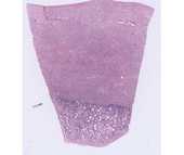
#66 Uterus Human progravid H&E
At low power identify the broad expanse of smooth muscle (the myometrium). Under higher power note that fascicles of smooth muscle are arranged in various planes. There are numerous blood vessels within the myometrium. The larger muscular arteries when cut in cross section appear as swirls of smooth muscle.
NOTE: Smooth muscle and connective tissue both stain pink with H&E. To distinguish between these two types of tissue special stains such as Mallory trichrome can be used. With the Mallory stain collagen stain blue and smooth muscle fibers stains.
Optional: #102 Small intestine (Bodian/silver)
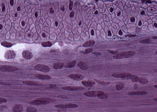 Find the two layers of smooth muscle. The basal lamina surrounding the muscle cells is stained with silver. Note that in the outer layer the muscle cells are cut in cross section. Examination of the silver-stained basal lamina at highest magnification reveals there are interruptions (i.e., the basal lamina is a dotted line). What is responsible for these discontinuities?
Comparison of skeletal and smooth muscle
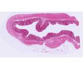
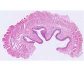
#32 Esophagus, Middle third, H&E
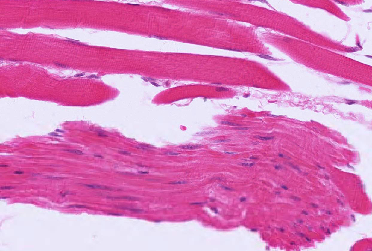 This is a good slide for comparing the morphological characteristics of smooth and skeletal muscles in both cross and longitudinal section, because these two types of muscle lie side by side in some areas.
This is a portion of a transverse section through the wall of the esophagus at or near the junction of its upper and middle segments. For orientation purposes, identify the following successive layers in the esophageal wall: (1) an inner lining of non- keratinized stratified squamous epithelium; (2) an adjacent loose connective tissue layer; (3) a layer formed by smooth muscle fibers (which have been cut in cross-section); (4) a subjacent connective tissue layer in which lie large blood vessels; (5) smooth and skeletal muscle fibers cut in longitudinal or oblique section, and (6) an outer layer rich in adipose cells and blood vessels.
Using higher magnification, examine the muscle layers and notice that the musculature is skeletal mixed with bundles of smooth muscle fibers due to the level of the esophagus at which this section was taken. In the skeletal muscle note the strong acidophilia and peripheral nuclei. In longitudinal sections identify cross-striations. In cross-section note that the skeletal muscle fibers resemble polygons separated by the endomysium with the nuclei are clearly visible at the periphery.
