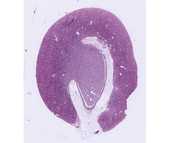
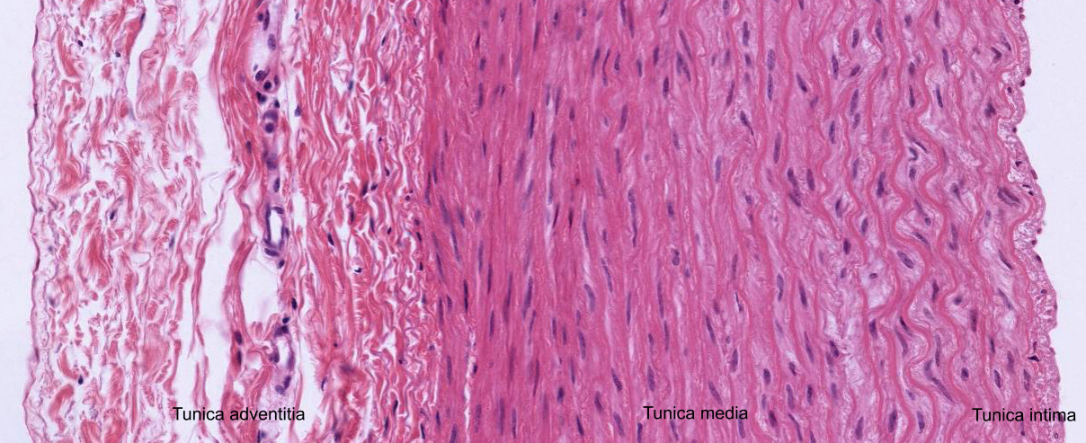
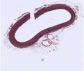
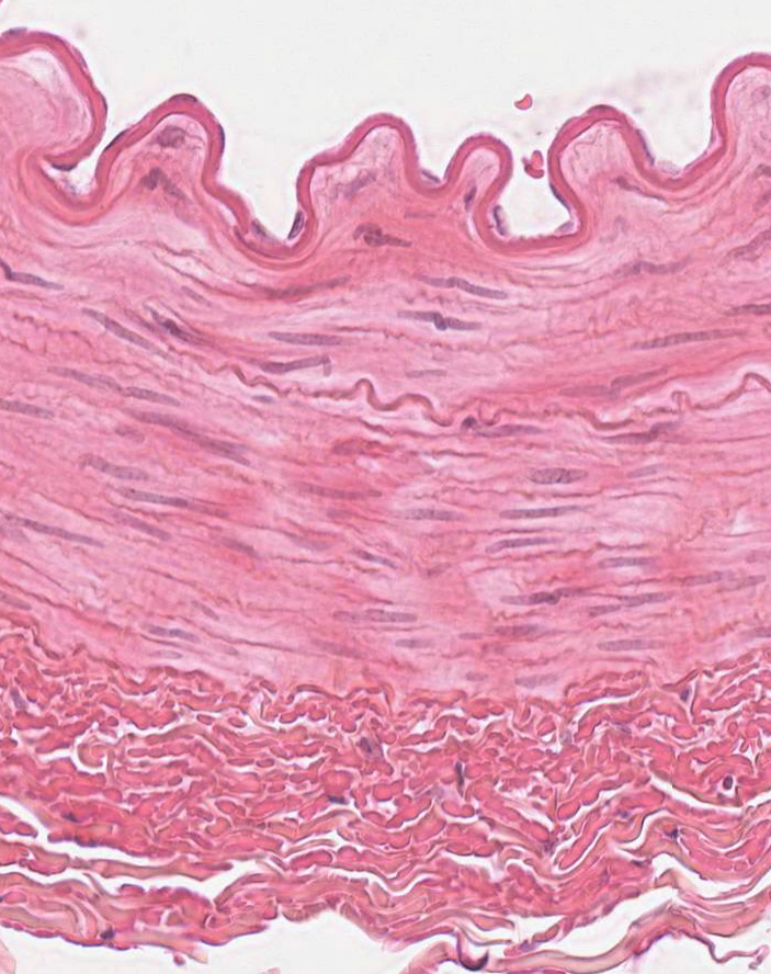
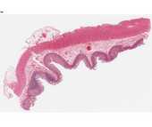
The Aorta
The sections on these slides are stained to demonstrate elastin, collagen and the cellular organization of the aorta. The aorta is an elastic artery which has a relatively thick tunica intima bounded by endothelium and the internal elastic membrane. The internal elastic membrane, however, is less obvious here than in the smaller muscular arteries. In the tunica intima smooth muscle cells run parallel to the long axis of the aorta while in the tunica media smooth muscle is spirally arranged. Within the tunica media the distribution of elastin in the elastic laminae is revealed as red-staining or black-staining material by the elastin stain. Elastin is not stained in the Masson preparations, but can still be seen as clear, refractile material surrounded by blue-staining collagen fibers. Both elastin and collagen are produced by smooth muscle cells, which are the only cell type within the tunica media.
#16 Aorta, Rhesus monkey, Cross Section
In addition to locating blood vessels, also observe the numerous sectioned nerves and adipose tissue. Note the presence of brown fat cells with their multilocular appearance.
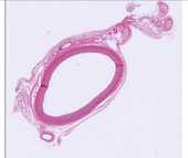
#20 Aorta, Cross Section (Elastin Stain)
In slides #16 and #20 the blood vessels of the aorta, the vasa vasorum, should be identified in the tunica adventitia.
In addition to locating blood vessels, also observe the numerous sectioned nerves and adipose tissue. Note the presence of brown fat cells with their multilocular appearance.
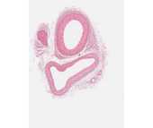
Arteries and Veins
Medium and small size arteries and veins occur in most of your slides, and they will be seen during the study of every tissue and organ. The following slides are particularly useful for distinguishing arteries and veins.
#113 Artery, vein, nerve, primate (H&E)
 Locate the cross section of the muscular artery, which can be identified by its thick wall and the scalloped luminal surface. The major component of the wall of the artery is spirally arranged smooth muscle (therefore seen here in longitudinal section). Examine the smooth muscle cells at high power. Note that the nuclei are elongated and that due to contraction of the vessel wall, some of them appear corkscrew shaped. The nuclei are relatively euchromatic (as compared to those of fibroblasts in the adventitia of the vessel). The smooth muscle cells, in addition to contracting to control the diameter of the vessel, also produce collagen and elastic fiber components of the muscular part of the vessel wall.
Identify large and small veins and venules, large and small arteries and arterioles. Compare the tunica media and the tunica adventitia in these vessel types. Note that in arteries and arterioles there is an internal elastic membrane. This appears wavy due to the contraction of the smooth muscle that underlies it. Identify capillaries and know their distinguishing characteristics.
Use any (or all) of the following slides to distinguish arteries, arterioles, veins and capillaries.
#117 Small Intestine
Use either of these slides to distinguish arteries, arterioles, veins and capillaries.
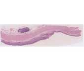
#4 Skin
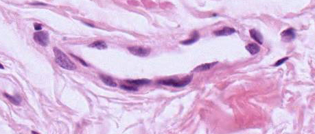 Note the presence of small blood vessels and sweat glands with ducts in the dermis. Blood vessels have an epithelium, whereas the sweat glands and ducts are lined by cuboidal epithelium.
#50 Kidney
Capillaries can easily be distinguished in the white fat in the renal pelvis.