Electron Micrographs
Examine the electron micrographs so that you understand the ultrastructural equivalents of the structures you have seen under the microscope.
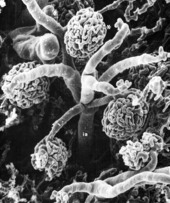
Vascular Arrangement in Glomeruli (SEM)
Scanning electron micrograph of arterial blood supply to renal glomeruli (*). The afferent arterioles (aa) that supply the glomeruli (asterisk) receive blood from an interlobular artery (ia).
Cormack, D.H. Ham's Histology, 9th ed., Lippincott, Philadelphia, 1987, p. 585.
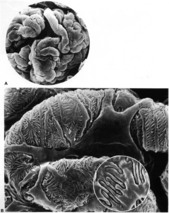
Glomerular Loops (SEM)
The glomerular visceral epithelium consists of podocytes that have primary (1) extensions that branch to form secondary (2) processes from which the podocyte feet (pedicels) extend as side branches and contact the glomerular basement membrane. Arrows indicate interdigitating secondary processes of two neighboring podocytes (Inset).
Cormack, D.H. Ham's Histology, 9th ed., Lippincott, Philadelphia, 1987, p. 578.
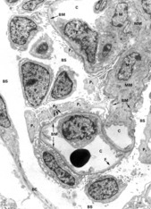
Bowman's Capsule and Glomerulus
Portion of a Bowman's capsule (BC) showing the parietal epithelium (BC) and Bowman's space (BS). The glomerular capillaries (C ) are lined by fenestrated (F) endothelium and surrounded by thick basement membrane (BM). The visceral epithelium that covers the capillaries is made up of podocytes (P) that extend branching foot processes (P1, P2) to lie on the basement membrane (BM). Mesangial cells (M) are found within the mesangial matrix (mesangium, MM).
Young, B & Heath, JW., Wheater's Functional Histology, 4th ed., Churchill Livingstone, London, 2000, p. 296.
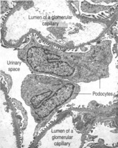
Glomerulus
Portion of glomerulus showing capillary loops covered by foot processes of podocytes.
Kierszenbaum, AL Histology and Cell Biology 2nd ed., Mosby Elsevier, 2007, p. 406.
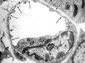
Glomerular Basement Membrane
Portion of glomerulus with three profiles of capillaries (C ). Endothelial lining is highly fenestrated (F). Podocytes (P) extend primary processes (P1) that give rise to numerous foot processes (P2) which lie on the thick basement membrane (BM). Adjacent foot processes( aka pedicels),(separated by filtration slits (FS) are derived from different podocytes. The basement membrane is a product of both the endothelial cells and podocytes.
Young, B & Heath, JW, Wheater's Functional Histology, 4th ed., Churchill Livingstone, London, 2000, p. 297.
Young, B & Heath, JW, Wheater's Functional Histology, 4th ed., Churchill Livingstone, London, 2000, p. 297.
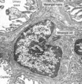
Mesangium
Mesangial cell and mesangial matrix. Mesangial cells are specialized pericytes with characteristics of smooth muscle cells and macrophages.
Kierszenbaum, AL Histology and Cell Biology 2nd ed., Mosby Elsevier, 2007, p. 406.
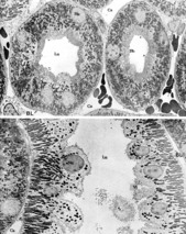
Portions of kidney tubule
Proximal (upper image) and distal (lower image) portions of the kidney tubule have different characteristics. The cells of the proximal tubule have a thick brush border (Bb), many mitochondria (Mi) and indistinct lateral borders. Cells in distal tubule (here the thick ascending limb) have a modest microvillous border and mitochondria are concentrated at the base. These cells are paler and bulge into the lumen (Lu). Their lateral borders are also indistinct due to lateral infoldings. Capillaries (Ca) abound in all regions.
Rhodin JAG, An Atlas of Ultrastructure, WB Saunders, Philadelphia, 1963, p. 97
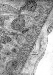
Basal Lamina of Proximal Tubule
Proximal tubule kidney (guinea pig). Basal membrane is highly infolded. Note fenestrated capillary at right.
Fawcett DW, The Cell: An Atlas of Fine Structure, WB Saunders, Philadelphia, 1966, p. 357.
Fawcett DW, The Cell: An Atlas of Fine Structure, WB Saunders, Philadelphia, 1966, p. 357.
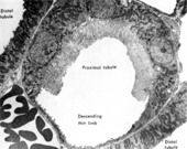
Kidney Tubules
Slightly oblique section through abrupt transition of thick descending limb with the thin loop of Henle. The brush border ends as the epithelium becomes squamous. Note portions of distal tubule and blood vessel with erythrocytes at left.
Bloom, W. and Fawcett, D.W. A Textbook of Histology 12th ed., W.B. Saunders, Philadelphia, 1994, p. 745
Bloom, W. and Fawcett, D.W. A Textbook of Histology 12th ed., W.B. Saunders, Philadelphia, 1994, p. 745
Glomerulus
Fenestrated capillary in nonglomerular region of kidney. Fenestrated capillary (in nonglomerular region of kidney). Arrows indicate fenestrae closed by diaphragms. In this cell the Golgi complex (G), nucleus (N), and centrioles (C ) can be seen. Note the continuous basal lamina on the outer surface of the endothelial cell (double arrows). Basal infoldings of kidney tubule epithelium are seen at upper right.
Junqueira, LC and Carneiro, J, Basic Histology 11th ed., McGraw-Hill, New York, 2005. p. 216.
Junqueira, LC and Carneiro, J, Basic Histology 11th ed., McGraw-Hill, New York, 2005. p. 216.
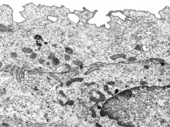
Transitional Epithelium (urothelium)
Superficial layer of transitional epithelium, showing the cytoplasm only of one epithelial cell. The plasma membrane contains plaques that are hinged such that when the bladder is contracted, peripheral membrane is internalized and can be everted when the bladder is distended.
Bloom, W. and Fawcett, D.W. A Textbook of Histology 12th ed., W.B. Saunders, Philadelphia, 1994, p. 761.
Bloom, W. and Fawcett, D.W. A Textbook of Histology 12th ed., W.B. Saunders, Philadelphia, 1994, p. 761.
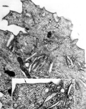
Transitional Epithelium (urothelium)
Luminal border of superficial epithelial cell of urinary bladder in the contracted state. Rigid plaques on the cell membrane, with hinge regions between them, give the luminal surface a spiky appearance and enable this part of the cell membrane to fold up into the cell. These internalized structures form fusiform vesicles (fv). Arrows indicate rigid plaques. Cormack, D.H. Ham's Histology, 9th ed., Lippincott, Philadelphia, 1987, p. 588.
