Pituitary Gland
The pituitary gland is a dual gland consisting of an epithelial component called the adenohypophysis and a neural component called the neurohypophysis. The adenohypophysis is derived from an outgrowth of oral ectoderm known as Rathke's pouch. It has three parts, thepars distalis (anterior lobe), pars tuberalis (enveloping the infundibular stalk), and pars intermedia (rudimentary in adults). The neurohypophysis is a neuroectodermal downgrowth from the floor of the diencephalon (part of the central nervous system) and includes the pars nervosa (posterior lobe) and the infundibulum.
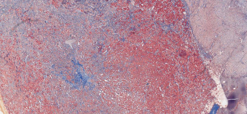 Each of the human pituitary slides in your collection has been stained with a different set of dyes. Identify the pars distalis, neurohypophysis, and remnants of the pars intermedia with the aid of your text. Under higher magnification, recognize glandular cells in the anterior lobe of each preparation. Note the arrangement of the hormone-secreting cells along the fenestrated sinusoidal capillaries.
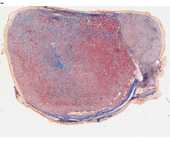
#73 Pituitary, (Masson's trichrome)
With Masson's stain, the acidophils are red and the basophils are blue. Chromophobes will be light orange or faded to grey. Note that red blood cells may be anything from red to blue. The cords and clumps of epithelial cells are sharply outlined by the blue collagenous fibers.
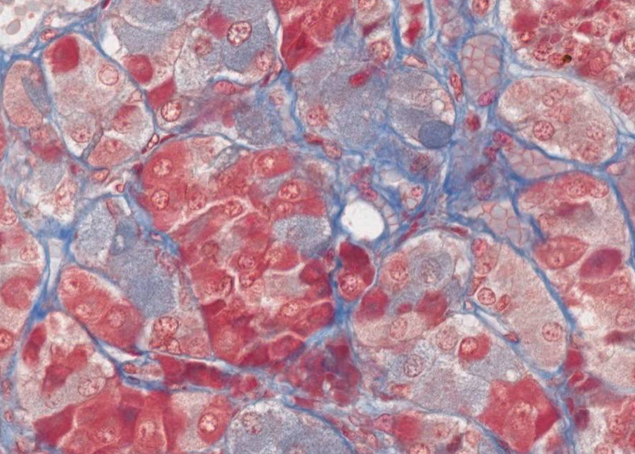 Note: When using Masson's trichrome or chrome alum-hematoxylin phloxin, the terms acidophils and basophils do not have the same meaning as when you use hematoxylin-eosin. The colors are dependent on the contents of the secretory granules. Acidophils synthesize proteins including growth hormone (GH) and prolactin. The basophils synthesize glycoproteins including luteinizing hormone (LH), follicle stimulating hormone (FSH), thyroid stimulating hormone (TSH) and adrenal cortical stimulating hormone (ACTH). Each of the hormones is made by a different cell, except the gonadotropins (LH and FSH), which may be made by the same cell. Chromophobes are thought to be cells in which the granules are depleted or are ACTH producing cells. The neuropil of the neurohypophysis is grayish and vacuolated. The red-pink nuclei belong to pituicytes (glial cells) and endothelia.
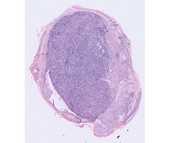
#74 Pituitary, H&E
The red or blue staining of the secretory granules is due to the acidophilia or basophilia of the hormone contained in the granules. The pars intermedia, which is not seen clearly on this slide, forms a cap around the neurohypophysis and separates it from the pars distalis. What kinds of cells are in the neurohypophysis?
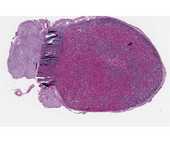
#75 Pituitary, (Chrome-hematoxylin and Phloxin)
Nuclei are purple-black, acidophils bright pink, basophils blue-black, chromophobes light blue to colorless. Note the regional variations in the distribution of the various cell types. Note also that in this pituitary there is a large colloid cyst (Rathke’s pouch).

Review the various hormones secreted by the basophils and acidophils (as defined in the trichrome stains) of the pars distalis.
This preparation demonstrates the Herring bodies (large magenta-stained swellings on the neurosecretory axons) in the neural lobe. What do Herring bodies represent? What hormones might you expect to find in these structures?
Where are the hormones of the neurohypophysis synthesized?
