Placenta
The placenta may be defined as an apposition or fusion of the fetal membranes with the uterine mucosa for the purpose of physiological exchange. The period of placentation is initiated by the attachment of the blastocyst to the endometrium, and it is terminated by the delivery of the newborn infant at the time of parturition. The placenta is the first organ to be differentiated, and performs functions analogous to those of the lung (gas exchange), intestine (nutrient absorption), kidney (excretion and ion regulation), liver (synthesis of serum proteins, steroid metabolism), pituitary (synthesis of hormones including gonadotropic and prolactin-like hormones), and gonads (incomplete synthesis of progestins and estrogens).
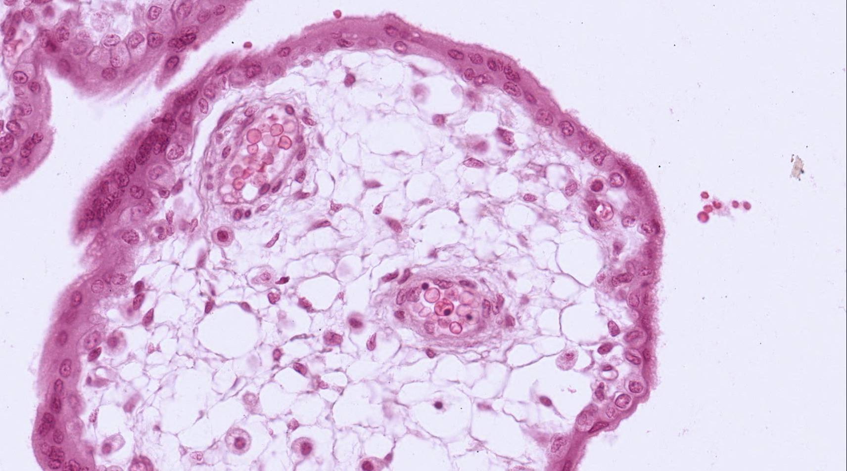
#98 Placenta, 2.5 months
Only the fetal surface of the placenta is present on this slide, so that the attachment of the fetal villi to the uterus cannot be studied. The fetal portion of the placenta consists of the chorionic plate, composed of an outer layer of trophoblast and an inner layer of vascularized extra-embryonic mesodermal connective tissue. The bulk of the placenta fetalis consists of outgrowths of villi from the surface of the chorionic plate. The villi are sectioned in many different planes, and their attachment to the chorionic plate may not be evident. Attached to the inner (fetal) surface of the chorionic plate is the amnion, consisting of an inner squamous amniotic epithelium and an outer layer of avascular mesoderm.
Study the chorionic villi in detail, and identify all of the layers that separate the maternal and fetal blood. These are:
1. syncytiotrophoblast (outermost layer, derived from cytotrophoblast; lines maternal blood space)
2. cytotrophoblast, which may be reduced in places
3. basement membrane of the trophoblast
4. fetal connective tissue, which may be greatly reduced in the region of apposition of the underlying fetal capillaries to the trophoblast
5. fetal capillary endothelium and its basement membrane.
Gases, nutrients, metabolites and other substances must pass through these layers to move from one circulation to the other. In life maternal blood fills the intervillous space, but it is generally washed out during tissue preparation.
Mitotic figures are occasionally seen in the cytotrophoblast, but not in the syncytiotrophoblast. Note the loose appearance of the cells forming the cores of the villi, and compare this with the condition in the villi at 6 months gestational age. Occasional nucleated fetal red blood cells, characteristic of earlier stages, can still be observed in the fetal vessels of the villi.
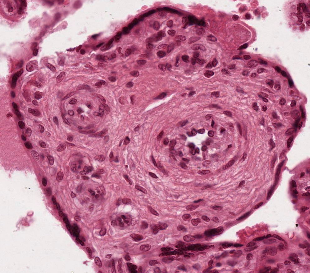
#100 Placenta, 6 months
 Considerable branching and diminution of the chorionic villi has occurred. Note the abundance and location of the fetal capillaries, the sparsity of the cytotrophoblast, and the nature of the syncytiotrophoblast. There are portions of syncytiotrophoblast (syncytial knots) floating free in the maternal blood. There is no endometrial tissue in this slide.
Be certain that you know the layers that form the separation between fetal and maternal blood in the placenta.
What is the placental source of human chorionic gonadotrophin, (HCG)?
What fluid bathes chorionic villi?

