Esophagus
As in both the oral cavity and in the pharynx, the mucosal surface of the esophagus is lined by stratified squamous epithelium that is non-keratinized in humans. In herbivores, the esophagus has a keratinized epithelium. The well-developed muscularis externa and the stratified squamous epithelial lining are well adapted for the rapid transport of food from the pharynx to the stomach. Diagnostic features of the esophagus are the combination of stratified squamous surface epithelium and the considerable thickness of the muscularis mucosae (up to 0.2 - 0.4 mm thick). The upper third of the muscularis externa contains mostly skeletal muscle, the middle third contains a mixture of skeletal and smooth, and the lower third contains only smooth muscle.
#32 Esophagus (and trachea), middle third, Human, (H&E)
Examine the wall of the esophagus starting with the stratified squamous non-keratinized epithelium. Underlying the epithelium is a layer of loose connective tissue and the muscularis mucosae. Note the intermingling of both skeletal and smooth muscle in the muscularis externa. Use this slide to review the histology of smooth and skeletal muscle, comparing them with the adjacent connective tissue of both the lamina propria and the submucosa. Also, identify the various types of vessels within the submucosa, as well as other layers.
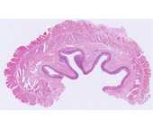
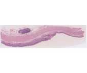
#33 Junction of Esophagus and Stomach, Human (H&E)
Locate the abrupt transition between the stratified squamous epithelium of the esophagus and the simple columnar epithelium of the stomach. Compare the muscle of the muscularis externa of the esophagus with that of the previous esophageal slides. How can you diagnose whether you are looking at the upper or lower portion of the esophagus? Note both the diffuse infiltration of lymphocytes and the scattered lymph nodules within the lamina propria of the stomach. A more detailed study of the various cell types of the gastric epithelium is considered on the next slide.