Oral Cavity
The epithelial lining of the oral cavity is of the stratified squamous type. In contrast to the skin it is non-keratinized. The major salivary glands arise as invaginations of the oral epithelium during the second month of embryonic development, and they are involved with the secretion of the watery, mucus, and enzymatic content of saliva. A description of these glands can be found in the following lab.
Tongue
The tongue is easily recognized because of its interlacing bundles of skeletal muscle that are disposed in three plane, and by its covering of stratified squamous epithelium that is elevated on the dorsal surface of the tongue into papillae. There are three types of papillae in humans, filiform, fungiform, and circumvallate.
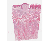
#116 Tongue, human, circumvallate papillae, H & E
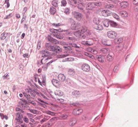 The lingual papillae are vertical elevations or projections of the epithelial surface and they contain a core of connective tissue derived from the lamina propria. In this section there are a number of filiform papillae and a single circumvallate papilla. Note the numerous taste buds on the lateral walls of the circumvallate papilla. Some of these have been sectioned through the taste pore. The three types of cell within the taste bud are sensory, supporting, and basal, but you should not attempt to distinguish them. Find a taste bud in which the taste pore is the plane of the section.
There are massive serous glands (glands of von Ebner, a.k.a. lingual salivary glands). In some sections the ducts of these glands may be seen to drain into the furrow of the circumvallate papilla. Note the bands skeletal muscle (artifactually separated) and identify blood vessels and nerves. There is considerable lymphatic invasion, particularly around the secretory portions of the serous glands.