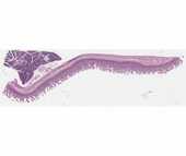
Small Intestine
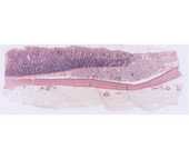
#35 Stomach and Duodenum, (H&E)
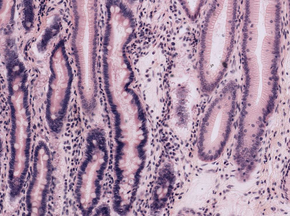 With low power, locate the junction of the stomach and duodenum. Look for differences in the epithelial surface and note the thickening of the muscularis externa of the stomach as it becomes the pyloric sphincter. Although the gastric mucosa is characterized by surface pits and the intestinal mucosa is characterized by finger-like villi, this distinction is not always readily apparent on sections. One of the best ways to distinguish between the two organs is to examine the surface epithelium that lines the pits or villi. In the stomach the cells all have a uniform appearance, since they are all mucus-secreting cells. In the intestinal villi however, most of the cells are absorptive cells, and interspersed between these are the characteristic mucous-secreting goblet cells. Goblet cells of the intestine will stand out when the slide is scanned under low power. In addition, a brush border can sometimes be seen on the free surface of the absorptive cells in well-preserved intestinal villi. To what ultrastructural feature does the brush border correspond? The duodenum is also characterized by the presence of mucous-secreting duodenal glands (of Brunner) in its submucosa.
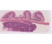
#36 Small intestine, duodenum, H&E
Identify the three components of the mucosa: epithelium, lamina propria and muscularis mucosae. Circular folds called plicae circulares are visible at low power on this slide. They further increase surface area for absorption. Note the submucosal glands of Bruner. The two layers of the muscularis externa are present and outside these is the adventitia. Between the two layers of the muscularis externa, identify elements of the myenteric plexus. The pancreas can be seen beneath the muscularis externa. This will be studied in the next lab.
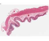
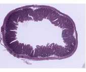
#117 Small Intestine, H&E.
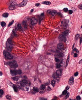 Identify the components of the wall of the small intestine. Note the presence of diffuse lymphatic tissue in the mucosa (GALT). In the ileum there are accumulations of lymph nodules called Peyer's patches. At the base of the intestinal crypts Paneth cells are found. These are characterized by the accumulation of large acidophilic granules in their apical cytoplasm, and by their strongly basophilic basal cytoplasm. The appearance of these cells is characteristic of enzyme-secreting cells. These cells secrete the anti-bacterial enzyme lysozyme.
#102 Small Intestine. (Bodian/silver)
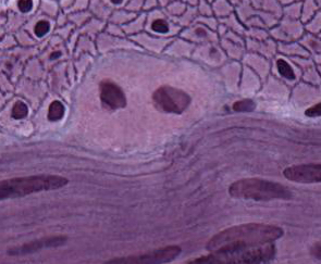 In addition, the Bodian silver stain demonstrates the enteroendocrine cells. These cells have granules along the basal surface, which secrete hormones into capillaries. The highest magnification will be required to find them. Note the basal lamina around smooth muscle cells in both longitudinal and cross-section. Myenteric neurons are also very nicely demonstrated in this slide.
#101 Small Intestine. (PAS and hematoxylin)
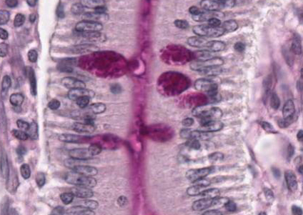 This slide is useful for demonstrating structures that contain glycosaminoglycans. The glycocalyx that covers the microvillus border of villus absorptive cells can be seen. Within the crypts of Lieberkuhn, which contain undifferentiated cells, the glycocalyx is not present. Note also the staining in the loose connective tissue of the lamina propria and the intense staining of goblet cells in the epithelium. In the lamina propria there are macrophages with irregular-sized PAS-positive inclusions. There are also mast cells, which are smaller than macrophages and filled with intensely stained granules. They are not usually found in the apical region of the villus. What accounts for the intense PAS-positivity of these cells?
