Major Salivary Glands
The major salivary glands (as well as the pancreas) are examples of exocrine glands, meaning the products of secretory cells are delivered via ducts to their functional site. These glands are classified according to: (1) the organization of the cells in the secretory portion of the gland e.g. tubular, alveolar (synonymous with acinar) or tubulo-alveolar (both types are present), and (2) the configuration of the cells that form the excretory duct (or ducts). If the duct is unbranched, the gland is called a simple gland. If however, the duct branches the gland is known as a compound gland.
 The parotid, submandibular, and sublingual glands are all compound (branched), tubulo-alveolar (acinar) glands with a merocrine (exocytotic) type of secretion. The secretory product is either serous (a protein product secreted in vesicles) or mucous (a large sulfated glycoprotein). Be sure that you can distinguish between serous and mucous cells. Serous cells have rounded, central nuclei and basophilic cytoplasm. The cytoplasm of mucous cells appears unstained due to the loss of the mucus product during tissue preparation. The nuclei of mucus cells are flattened at the base of the cells. There are myoepithelial cells between the basal lamina and the basal plasma membrane of the secretory cells.
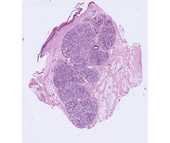
#40 Parotid gland, (H&E)
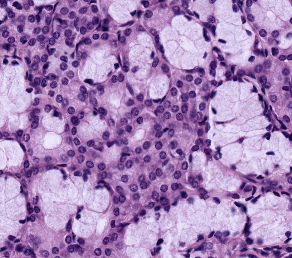
 This gland is purely serous in the adult human, so that all of the secretory units or acini have a similar appearance. A fibrous connective tissue capsule surrounds the gland and sends septa inward that subdivide the gland into lobules.
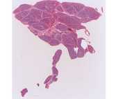
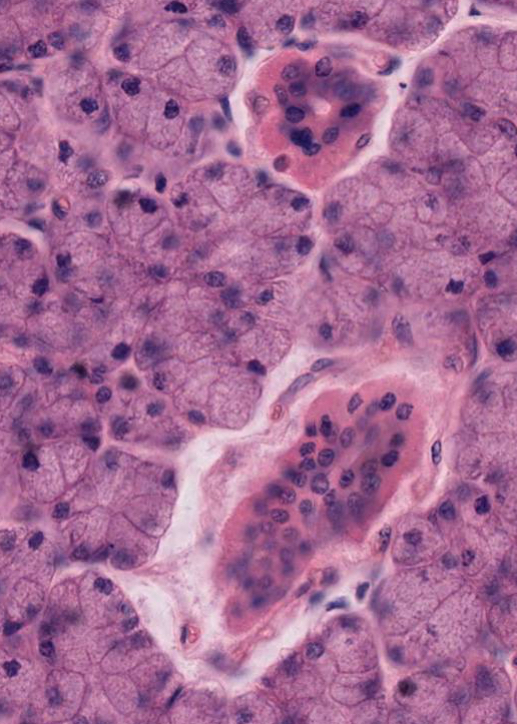
Scan the slide on low power and observe that within each lobule there are several prominent ducts with a more distinct lumen; these stand out sharply from the surrounding acini because of their acidophilia. These are the intralobular or striated ducts. Larger interlobular or excretory ducts are embedded in the extensive interlobular connective tissue, and their epithelium may be either simple columnar, pseudostratified, or stratified. Goblet cells are occasionally seen in the interlobular ducts. The secretory acinus is connected to the intralobular duct by a thin intercalated duct, which is difficult to see in this slide. These intercalated ducts consist of a cuboidal epithelium, and their diameter is less than that of either the acinus or the intralobular duct.
#41 Sublingual Gland, (H&E)
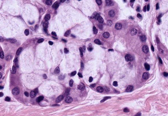 This is a mixed gland, with both mucous and serous secretory units. Most of the acini are either purely mucous or are mixed. The mixed secretory units consist of a mucous acinus capped by a crescent-shaped aggregation of serous cells called a serous demilune.
The submandibular gland is also a mixed gland that is predominantly serous.